


A research team led by Professor Shan Chongxin from the School of Physics at Zhengzhou University has developed a novel "resonance afterglow" amplification strategy using carbon nanodot polymer composites. The work, published in the journal Nature Communications, offers a new approach to ultrasound-responsive nanoprobes and tumor imaging-guided surgery.
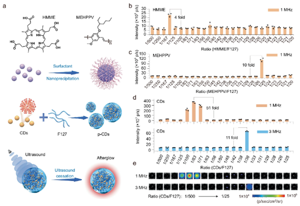
Preparation of ultrasound-responsive afterglow matrixes with different fluorophores. [Photo/zzu.edu.cn]
Afterglow imaging can suppress tissue autofluorescence, making it suitable for high-contrast bioimaging. However, traditional light-excited afterglow materials struggle to simultaneously achieve a long afterglow duration, high intensity, and deep activation. Ultrasound offers advantages as a non-optical excitation source, but existing systems suffer from low energy conversion efficiency.
The team assembled near-infrared carbon nanodots with amphiphilic block polymers to form a micron-scale interconnected gel network. This structure resonates with 1 MHz ultrasound, amplifying afterglow intensity by up to 51 times. Finite element simulations confirmed an inverse relationship between assembly size and resonant frequency. The mechanism involves piezoelectric-induced reactive oxygen species and peroxide intermediate decomposition.
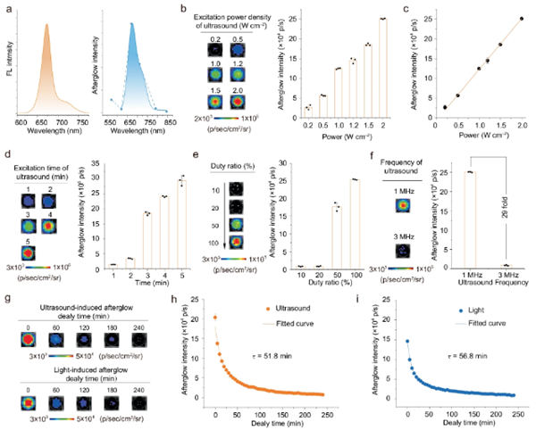
Ultrasound-responsive afterglow performance. [Photo/zzu.edu.cn]
A single two-minute ultrasound treatment produced in vitro afterglow lasting 240 minutes. The material achieved 2.5 cm tissue penetration, with in vivo afterglow signals exceeding 60 minutes in mice and a signal-to-noise ratio of 11.5, enabling tumor imaging and precise resection.
This work establishes a theoretical framework for size-frequency resonance, opening new pathways for next-generation ultrasound-responsive bioprobes and surgical navigation.
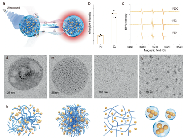
Mechanism of ultrasound-induced afterglow from p-CDs. [Photo/zzu.edu.cn]